Simple - 3-step deployment
Proven
Fast Sealing1
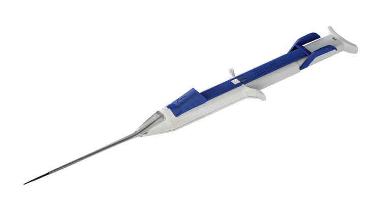
The FemoSeal™ Vascular Closure System is designed for mechanical closure of common femoral artery access sites following diagnostic or interventional catheterization procedures using a 7F (2.33 mm) or smaller procedural sheath.
FemoSeal™ provide active mechanical closure using a bioabsorbable seal, designed to promote rapid hemostasis without leaving permanent implants behind.
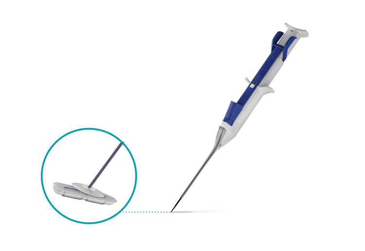
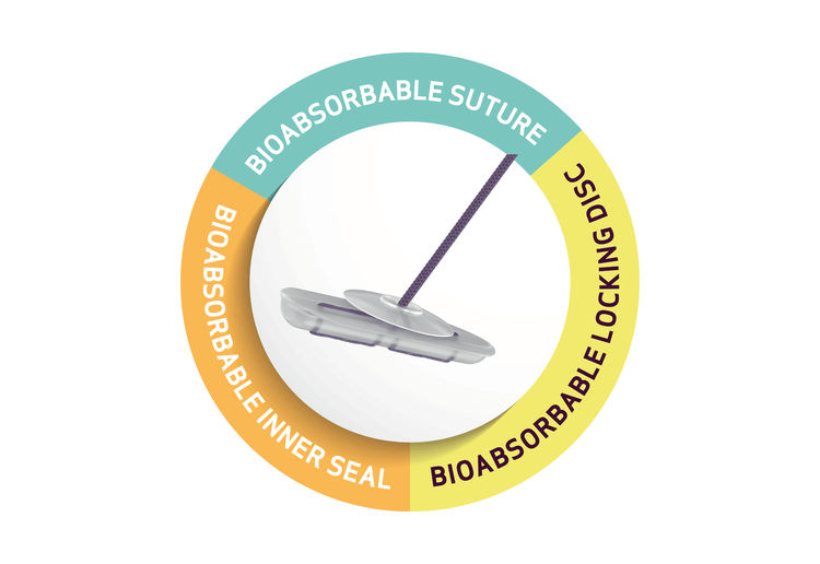
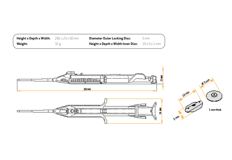
The FemoSeal™ Vascular Closure System promotes mechanical hemostasis by deploying implantable closure elements inside and outside the common femoral artery :
- Inner Sealing Disc: bioabsorbable polymer placed against the inside of the vessel wall
- Outer Locking Disc: bioabsorbable polymer placed on top of the arteriotomy in the tissue tract to "lock“ or maintain the seal at the end of the procedure
- Bioabsorbable Suture: binds inner sealing disc and outer locking disc together to form the seal
Watch the FemoSeal™ in action
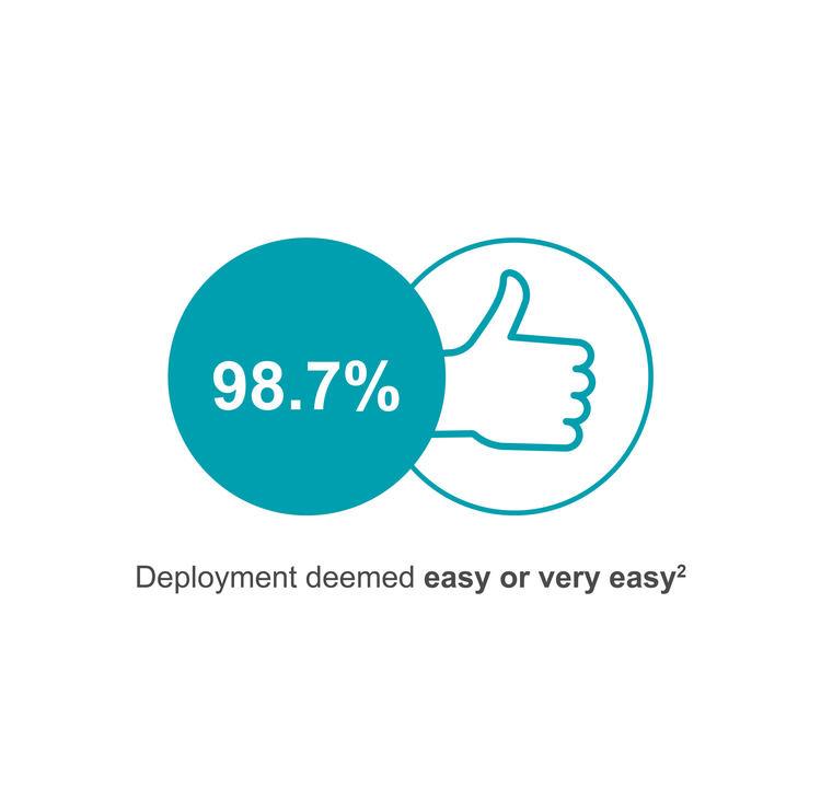
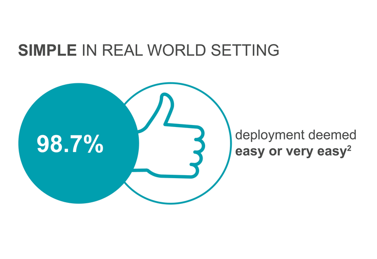
FemoSeal™ Vascular Closure System deployment is easy with only 3 steps:
- LOCATE the artery
- SET the discs
- SEAL the puncture
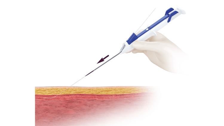
STEP 1 : LOCATE THE ARTERY*
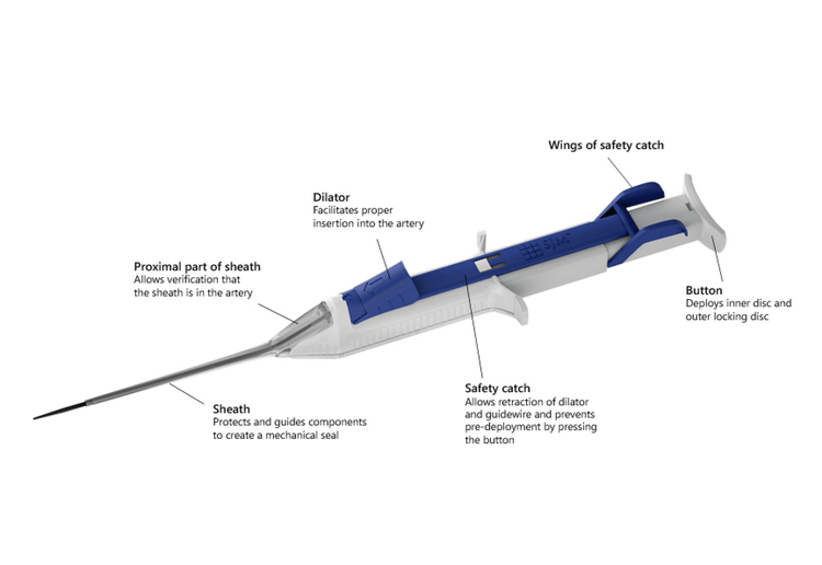
1. After assessing the puncture site by fluoroscopy or ultrasound, insert the dilator into FemoSeal™ Unit until they snap together and marked signs meet. Insert the guidewire through the procedural sheath. Remove the procedural sheath and hold the guidewire in position. Thread the FemoSeal™ over the guidewire and insert the sheath into the artery.
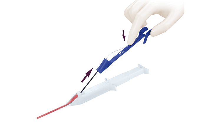
STEP 1 : LOCATE THE ARTERY*
2. Grip and squeeze the wings of FemoSeal™ Safety Catch, lift and retract the Safety Catch slowly along with the guidewire by 2-4 cm until completely removed. Blood should appear in the proximal part of the sheath, verifying that the sheath is in the artery.
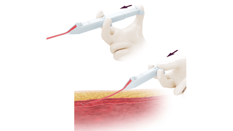
STEP 2 : SET THE DISCS
3. Deploy the inner seal by pressing down the button completely in one continuous movement. The inner seal establishes hemostasis.
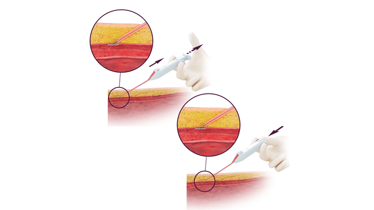
STEP 2 : SET THE DISCS
4. Pull back the FemoSeal™ and lift it with an angle (not less than 45°) until the depressed button springs back. The inner seal establishes hemostasis. Maintain suture tension while deploying the outer locking disc. Deploy the disc by completely pressing down the button again.
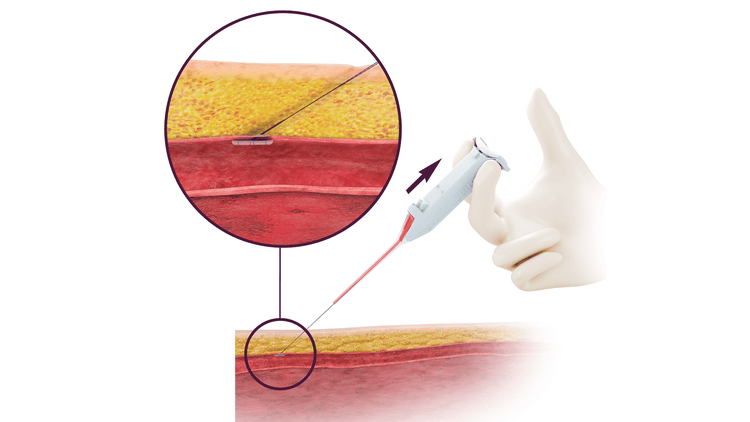
STEP 3: SEAL THE PUNCTURE
5. Remove the FemoSeal™ unit and verify hemostasis.
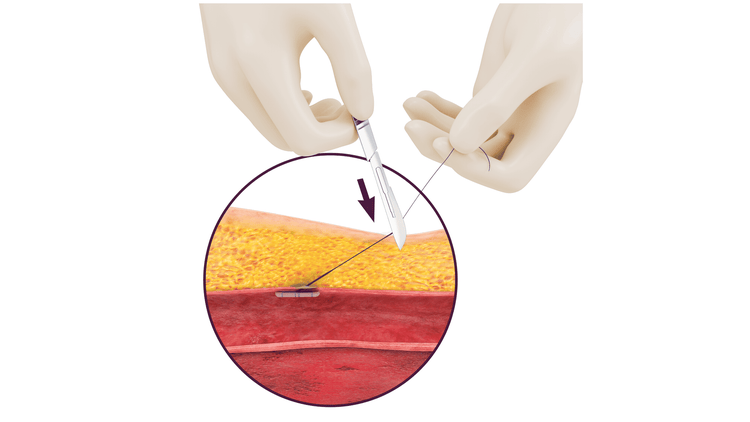
STEP 3: SEAL THE PUNCTURE
6. Cut the suture below skin level using a sterile instrument. Clean puncture site and apply a sterile dressing so that it can easily be observed during recovery.
SIMPLE – 3-step deployment
PROVEN
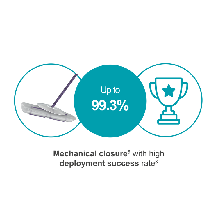
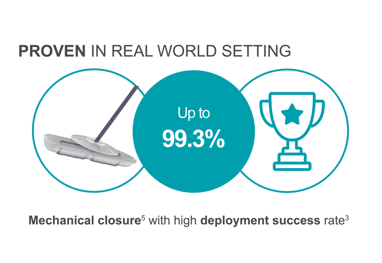
FemoSeal™ has been clinically proven to achieve
- High deployment success rates in interventional cardiology & peripheral interventions2,3
- Hemostasis without adjunctive manual compression or wound dressing2
- Superior safety outcomes compared to extravascular Vascular Closure Devices4
- Significantly lower rates of access-site related vascular complications vs manual compression4
FAST SEALING1
FemoSeal™ has been clinically proven to
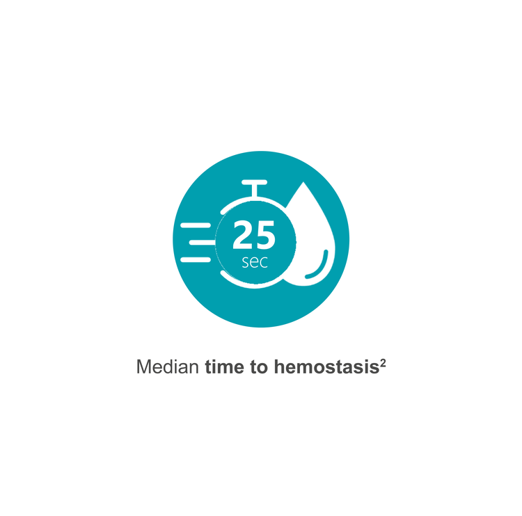
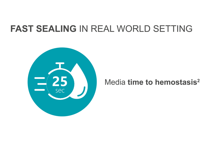
- Support median time to hemostasis of 25 seconds2
- Reduce time to hemostasis by 95% vs manual compression4
Troubleshooting Videos
Downloads
References
Disclaimer
Not all products are available for sale in all countries. This information is provided only in respect to markets where these products are approved or cleared. Please contact your Terumo local sales representative for more information. Refer to Instructions for Use for contraindications, warnings, precautions and additional information. All brand names are trademarks or registered trademarks owned by TERUMO CORPORATION, its affiliates, or unrelated third parties.
Didn't find your product?
Consult our products in our Product Index