Physician: Roberto Iezzi, MD
Location: Institute of Radiology - Bioimaging Department Agostino Gemelli Hospital Foundation - Catholic University - Rome – Italy
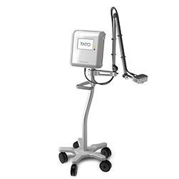
Featured product: TATO Thermal Ablation Treatments for Oncology
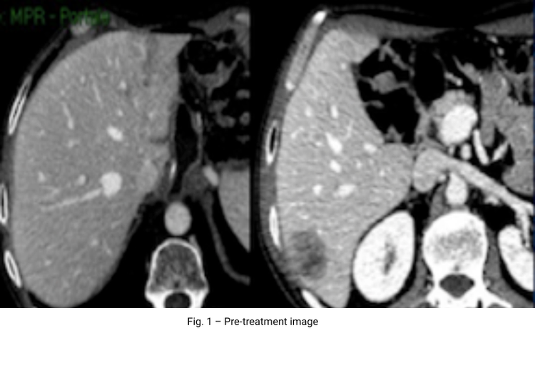
59 yo woman in follow-up after pancreatic resection from advanced-stage pancreatic ductal adenocarcinoma (two years prior the presentation). In the interim, she underwent wedge resection of lung lesions, cervical cortectomy and fusion (c-6 metastasis) and several lines of chemotherapy (gemcitabine-based). Abdominal contrast enhanced CT revealed two hepatic metastatic lesions: 1.4x2.4 cm metastasis in segment III and 2.6x4.8 cm metastasis in segment VI (Fig.1).
Laboratory data: hemoglobin (Hb) 12.2 g/dL; platelet count (PLT) 210,000/mm³; prothrombin time (PT) 14s; INR: 1.08; CA 19-9: 80.9 U/mL.
Based on lesion size, we decided to perform a single-step combined treatment of MWA followed by DEB-TACE for the large sVI metastasis and MWA only for the sIII lesion, in the same stage in angiosuite.
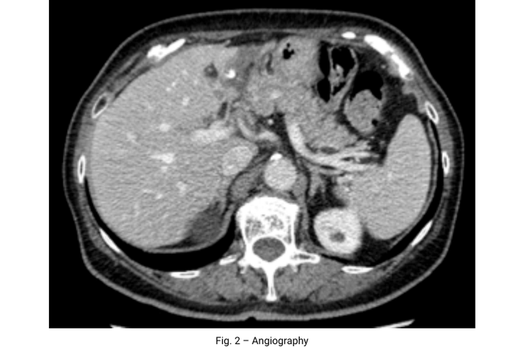
Procedure: Patient under sedation with fentanyl citrate (0.1–0.2 mg) and local anesthesia. Patient also received antibiotic prophylaxis and gastric protection. Hepatic angiography was performed using a right common femoral approach to map arterial tumor supply. Under US-guidance, a 14G uncooled TATO probe was introduced into the sVI nodule with 40 W of applied power in 10 minutes. During the same single session, a second 14G uncooled probe was inserted under US guidance in sIII lesion, with 30 W of applied power in 10 minutes. Immediate angiography revealed a reactive hyperaemia in sVI lesion (Fig 2); for this lesion, superselective transarterial chemoembolization using 100 μm drug-eluting embolics (LifePearl, Terumo) loaded with doxorubicin 50 mg was also performed using a coaxial technique and a 2.7-F microcatheter (Progreat, Terumo). The full planned dose was delivered and a complete tumour arterial devascularisation was achieved.
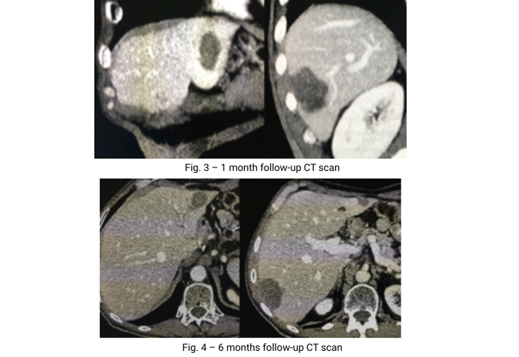
Patient tolerated the procedure well, without significant post-embolisation syndrome, and without reported complications; hospital discharge obtained 24 hours after procedure. 1-month CT follow up showed a complete response, with a treated area of 2.6x3.6 cm for sIII lesion and 3.8x5.6 cm for sVI lesion, without residual or recurrent vital tissue (Fig.3), as also confirmed by 6-month CT follow-up (Fig.4).
The possibility to handle up more applicators with a single generator allows interventional radiologists to operate more lesions in a single session. Furthermore, the coverage in Teflon makes the probe echogenic and non sticky allowing an easy and very high accurate positioning in the lesion, without risks and complications.